“Pink eye,” or conjunctivitis, is a non-specific finding that simply indicates conjunctival inflammation. The vast majority of children who present with “pink eye” will have a simple conjunctivitis. Neonatal conjunctivitis is an infection occurring during the first month of life, and is treated by administering antibiotics immediately after birth. Other causes of a “red, teary eye” in a newborn include congenital glaucoma and nasolacrimal duct obstruction. The most common causes for pediatric pink eye are allergic conjunctivitis, bacterial conjunctivitis, viral conjunctivitis, and blepharitis.
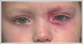
Bilateral Allergic Conjunctivitis.
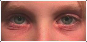
The patient has severe contact irritation of the lower lid skin secondary to ocular allergic conjunctivitis.
Note the marked redness on the conjunctiva in the patient.
Allergic Conjunctivitis
Allergic (hay fever) conjunctivitis is very common and affects approximately 10% of the general population. Seasonal allergic conjunctivitis produces itching and tearing and is usually associated with nasal congestion and other symptoms. For chronic, recurrent conjunctivitis, therapy consists of removal of environmental allergens and the use of topical eye drops that stabilize the cell reaction (mast cell stabilizers) to the allergen. It usually requires 2 to 3 days of continued use to reduce symptoms. Immediate relief can be obtained by the use of topical antihistamines. Topical corticosteroids are reserved for severe allergic conjunctivitis such as vernal conjunctivitis, and are only used for short courses of a few days.
Bacterial Conjunctivitis
The eye is constantly exposed to bacteria, but tear defense mechanisms work to prevent infection. When bacterial infections do occur, they present as watery irritation of the eyes that can progress to a thick mucous discharge. Children with a bacterial conjunctivitis often complain that their eyelids stick together in the morning. There is usually redness and crusting of the lashes. Antibiotics are often prescribed and used for 7 days.
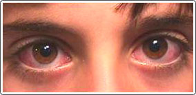
Viral Conjunctivitis
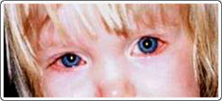
Child with H-flu and hemorrhagic conjunctivitis in the left eye. The bleeding usually clears up on its own in a couple of weeks.
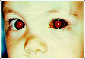
Chemical Conjunctivitis
Child with chemical conjunctivitis caused by contact with nail polish. The eye and skin cleared up with no lasting side effects.
In most cases, the key to preserving the integrity and health of the eye is immediate and ongoing rinsing of the eye with water for at least 5-10 minutes. Then, rapid transport to the doctor’s office or emergency room, if necessary, is warranted.
Blepharitis
Blepharitis, or eyelid inflammation, is one of the most common causes of pediatric pink eye. The two most common types of blepharitis are staphylococcal blepharitis and meibomian gland dysfunction. The best treatment for both is lid hygiene (baby shampoo lid scrubs) and topical antibiotics. Children with staphylococcus blepharitis complain of itching and burning and often awake with their eyelids stuck together with crusting. Their eyes are irritated, but there is not the “true” itching as with allergic conjunctivitis. Other signs include scales at the base of the eyelashes. Meibomian gland dysfunction produces irritation, burning, and redness of the eyelid margins and conjunctiva. Obstruction of the meibomian gland opening may result in an acute infection, but more commonly produces a chalazion. A chalazion appears as a lump near the eyelid margin, either on the upper or lower lid. Chalazia may resolve on their own over several weeks; however, applying hot soaks several times a day helps the drainage of lipid material. If it does not resolve, incision and drainage may be necessary.
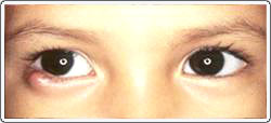
Child with bilateral blepharitis. Note the redness of the lid margins caused by irritation. There is tearing and some redness of the white part of the eye.
Sometimes blepharitis can lead to a chalazion that occurs when the lash opening at the skin becomes blocked and fills up with pus and excess debris.